
Fig. S5
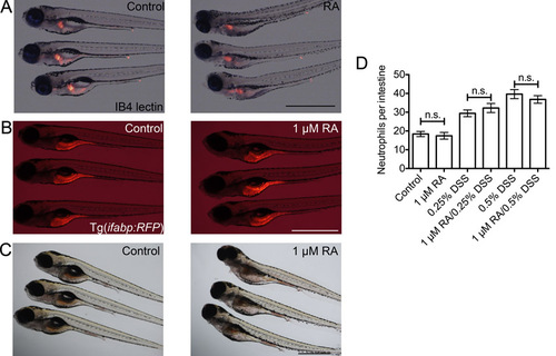
Doses of exogenous retinoic acid used in this study do not overtly affect intestinal patterning. (A) Representative images of whole mount IB4 lectin staining of the oesophageal-intestinal bulb junction in control and 1 μM RA-treated larvae. (B) Comparison of RFP expression in Tg(ifabp:RFP)as200 control and 1 μM RA-treated larvae. (C) Comparison of neutral red endocytosis by mid-intestinal epithelium in control and 1 μM RA-treated larvae. (D) Enumeration of intestinal neutrophils in larvae co-treated with 1 μM RA and DSS from 3 dpf (n≥30, two biological replicates). Error bars indicate S.E.M., P values determined by ANOVA. All scale bars indicate 1 mm. |