
Fig. 3
- ID
- ZDB-FIG-120717-12
- Publication
- Biran et al., 2012 - Neurokinin Bs and neurokinin B receptors in zebrafish-potential role in controlling fish reproduction
- Other Figures
- All Figure Page
- Back to All Figure Page
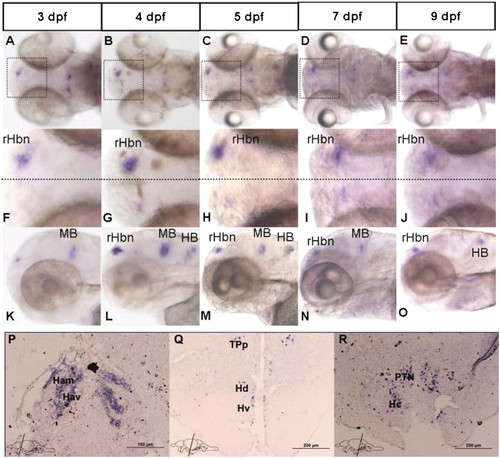
(A-O) Localization of tac3a during early stages of development, as detected by whole-mount ISH. tac3a is dominantly expressed in the right habenular nuclei, midbrain, and hindbrain. Dorsal view of larva heads, anterior is to the left (A-E). High magnification of boxed area in Upper panel. Note the unilateral expression of tac3a to right of the midline (dotted line) in the habenula (F-J), lateral view of larva heads (K-O). rHbn, right habenula; MB, midbrain; HB, hindbrain. (P-R) Localization of tac3a in adult zebrafish brain as indicated by ISH (nomenclature according to ref. 40). Tac3a mRNA-expressing cells were observed in the ventral (Hav) and medial (Ham) habenula (P) tac3a mRNA-expressing cells detected in the periventricular nucleus of posterior tuberculum (TPp), dorsal (Hd), and ventral zone (Hv) of periventricular hypothalamus (Q) tac3a mRNA-expressing cells observed in the posterior tuberal nucleus (PTN) and central zone (Hc) of periventricular hypothalamus (R). (Magnification: A-E and K-O, 40×; F-J, 120×). |
| Gene: | |
|---|---|
| Fish: | |
| Anatomical Terms: | |
| Stage Range: | Protruding-mouth to Adult |