
Fig. 1
- ID
- ZDB-FIG-120522-10
- Publication
- Barros-Becker et al., 2012 - Persistent oxytetracycline exposure induces an inflammatory process that improves regenerative capacity in zebrafish larvae
- Other Figures
- All Figure Page
- Back to All Figure Page
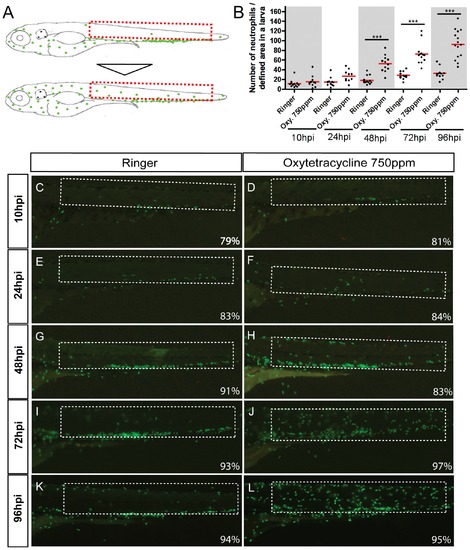
Exposure to oxytetracycline triggered an inflammation process. Incubation of Tg(mpx:GFP) transgenic larvae (that express GFP exclusively in neutrophils) in oxytetracycline 750 ppm induce a progressive migration of neutrophils from to the caudal hematopoietic tissue (CHT) to the entire tail. (A) Scheme of neutrophils localization in control and experimental larvae. (B) Quantification of neutrophils migration into the selected area. (C–L) Close up of the tail of control and experimental embryos at 10 hour post incubation (hpi), 24hpi, 48hpi, 72hpi and 96hpi. At 10hpi (C, D) and 24hpi (E, F) we did not detect any significant neutrophils migration. Later, at 48hpi (G, H) a discrete number of neutrophils migrate from de CHT to the tail. At 72hpi (I, J) and 96hpi (K, L), the presence of the GFP positive cells in the tail becomes clearly evident. The numbers of larvae that presented the phenotype shown is expressed as a percentage. For all experiments, at least 13 larvae were used for each condition. *** p<0.001. |