
Fig. 3
- ID
- ZDB-FIG-120426-24
- Publication
- Huang et al., 2012 - Novel expression patterns of metabotropic glutamate receptor 6 in the zebrafish nervous system
- Other Figures
- All Figure Page
- Back to All Figure Page
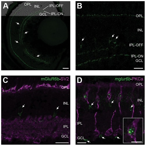
Subcellular localization of mGluR6b in the zebrafish retina. Z-projections of confocal image stacks of immunohistochemically labeled larval and adult retinal cross-sections. A: A larval retina at 5 dpf stained with the mGluR6b antibody shows labeling in the outer plexiform layer (OPL) and in an ON- and an OFF-layer of the inner plexiform layer (IPL). In addition, single cells adjacent to the IPL in the INL and the GCL (arrowheads) express mGluR6b as well. Scale bar = 20 μm. B: In the adult zebrafish retina, mGluR6b stains similar structures as in the larval retina with the exception that we could only detect labeled cells in the inner part of the INL (arrowhead) but not in the GCL. Scale bar = 15 μm. C: A doublelabeling of mGluR6b (green) and SV2 (magenta) in an adult retinal cross-section reveals the postsynaptic localization of mGluR6b in the OPL. Again, an mGluR6b expression in single cells of the proximal IPL is detected (arrows). Scale bar = 15 μm. D: Fluorescent in situ hybridization of mglur6b (green) combined with an immunohistochemical labeling of ON-bipolar cells by PKCalpha (magenta) confirms the localization of mGluR6b in ON-bipolar cells of the INL. Scale bar = 15 μm. Arrows point to cells of the proximal INL and the GCL expressing mglur6b. The insert shows a close up of an ON-bipolar cell body and its fluorescent mglur6b signal in the cytosol. Scale bar = 5 μm. |