
Fig. 7
- ID
- ZDB-FIG-120412-29
- Publication
- Idda et al., 2012 - Circadian timing of injury-induced cell proliferation in zebrafish
- Other Figures
- All Figure Page
- Back to All Figure Page
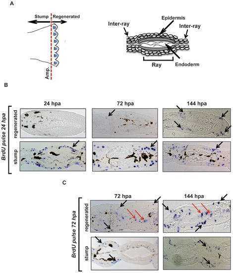
Early proliferating cells contribute to the formation of the new epidermis. (A) Left section: Schematic cartoon of an adult zebrafish caudal fin where the amputation site is indicated (Amp.) and the location of the stump and blastema (b) regions is defined. Right section: Schematic diagram of a transverse section through the zebrafish adult caudal fin. The identity of the principal structures is indicated. (B) Transverse sections of fins that 24 hours following amputation were labeled for 15 minutes with BrdU and then sampled at 24, 72 and 144 hpa. Histological sections through the tip of the new regenerating fin tissue (regenerated) and through the “original” portion of the fin (stump) are represented. Representative blue stained BrdU positive nuclei are indicated by black arrows and are predominantly restricted to the epidermal layers of the stump at all time points and in the regenerated epidermis at 72–144 hpa. (C) Sections from a comparable experiment to that presented in panel B, except that the 15 minutes BrdU labeling period was performed 72 hours after amputation. BrdU positive nuclei are visible in both epidermis (black arrows) and in the blastema region (red arrows) at all time points in the regenerating tissue. |