
FIGURE
Fig. S3
- ID
- ZDB-FIG-120316-76
- Publication
- Buck et al., 2012 - Ototoxin-induced cellular damage in neuromasts disrupts lateral line function in larval zebrafish
- Other Figures
- All Figure Page
- Back to All Figure Page
Fig. S3
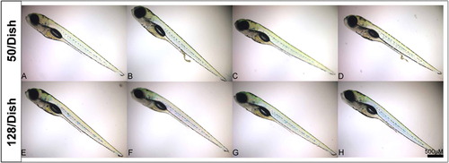
Development was unaffected by altering the density at which larvae are raised. (A-D) Representative images of larvae reared at a density of 50 per Petri dish (taken from a sample of 150 animals). (E-H) Representative images of larvae reared at a density of 128 per Petri dish (taken from a sample of 128 animals). Note normal development of eye, body pigmentation, body length, and swimbladder inflation for all animals. Scale bar = 500 μm. |
Expression Data
Expression Detail
Antibody Labeling
Phenotype Data
Phenotype Detail
Acknowledgments
This image is the copyrighted work of the attributed author or publisher, and
ZFIN has permission only to display this image to its users.
Additional permissions should be obtained from the applicable author or publisher of the image.
Reprinted from Hearing Research, 284(1-2), Buck, L.M., Winter, M.J., Redfern, W.S., and Whitfield, T.T., Ototoxin-induced cellular damage in neuromasts disrupts lateral line function in larval zebrafish, 67-81, Copyright (2012) with permission from Elsevier. Full text @ Hear. Res.