
FIGURE
Fig. 6
- ID
- ZDB-FIG-120315-5
- Publication
- Odenthal et al., 1996 - Mutations affecting xanthophore pigmentation in the zebrafish, Danio rerio
- Other Figures
- All Figure Page
- Back to All Figure Page
Fig. 6
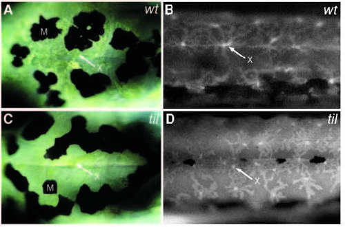
In tilsit mutants xanthophore pigment appears condensed whereas the cells appear more spread out. Dorsal view of day-6 larva in the head region posterior to the eyes, viewed with Nomarski optics (A,C), and lateral view of ammonia-induced fluorescence in the trunk under UV-light epi-illumination (B,D). (A,B) Wild type and (C,D) homozygous mutant larva of tilty130b. M, melanophores; X, and arrow, xanthophores. |
Expression Data
Expression Detail
Antibody Labeling
Phenotype Data
| Fish: | |
|---|---|
| Condition: | |
| Observed In: | |
| Stage: | Day 6 |
Phenotype Detail
Acknowledgments
This image is the copyrighted work of the attributed author or publisher, and
ZFIN has permission only to display this image to its users.
Additional permissions should be obtained from the applicable author or publisher of the image.
Full text @ Development