
Fig. 2
- ID
- ZDB-FIG-120216-42
- Publication
- Matsumoto et al., 2012 - Molecular cloning and expression of the col2a1a and col2a1b genes in the medaka, Oryziaslatipes
- Other Figures
- All Figure Page
- Back to All Figure Page
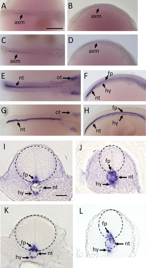
Expression patterns of col2a1a and col2a1b mRNAs in medaka embryo detected by whole-mount insitu hybridization at 1–2 dpf using the col2a1a probe (A, B, E, F, I, and J) and col2a1b probe (C, D, G, H, K, and L). Results at 1 dpf (A–D) and 2 dpf (E–L) in the dorsal view (A, C, E, and G) and lateral view (B, D, F, and H). (I–L) is a transverse section of the specimen at 2 dpf. (I and K) is the anterior notochord, (J and L) is the posterior notochord. The broken line of (I–L) is an outline of the neural tube and notochord. The sense probe showed no signals in any tissues through all of the stages tested (data not shown). axm, axial mesoderm; fp, floor plate; hy, hypochord; nt, notochord; ot, otic vesicle. Scale bar = 250 μm (A–H), 20 μm (I–L). |
Reprinted from Gene expression patterns : GEP, 12(1-2), Matsumoto, T., Deguchi, T., Kawasaki, T., Yuba, S., and Sato, J., Molecular cloning and expression of the col2a1a and col2a1b genes in the medaka, Oryziaslatipes, 46-52, Copyright (2012) with permission from Elsevier. Full text @ Gene Expr. Patterns