
Fig. 8
- ID
- ZDB-FIG-111213-28
- Publication
- Kasprick et al., 2011 - Microanatomy of adult zebrafish extraocular muscles
- Other Figures
- All Figure Page
- Back to All Figure Page
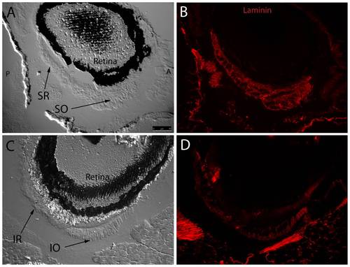
Fluorescent immunohistochemistry reveals an even distribution of laminin throughout myocyte basement membranes at the SR/SO and IR/IO insertion sites. DIC images show SR and SO overlapping near their globe insertion site on the superior side of the eye and IR and IO in a similar arrangement on the inferior side of the eye (A,C). Fluorescent overlays highlight uniform laminin (red) distribution throughout basement membranes of individual myocytes (B,D). Broader areas of laminin expression surrounding the muscle represent either broad laminin expression throughout a myotendinous junction or simply myocyte basement membrane cut in perfectly tangential section. Images were captured with a 20x objective lens. |