
Fig. 7
- ID
- ZDB-FIG-111202-27
- Publication
- Feng et al., 2010 - T-lymphoblastic lymphoma cells express high levels of BCL2, S1P1, and ICAM1, leading to a blockade of tumor cell intravasation
- Other Figures
- All Figure Page
- Back to All Figure Page
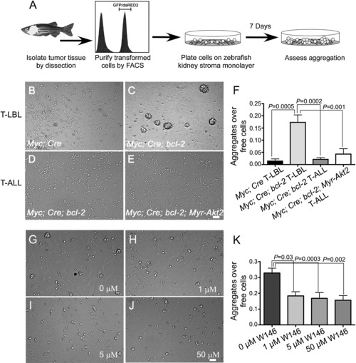
Bcl-2-Overexpressing T-LBL Cells Display Increased Aggregation that Can Be Overcome by Akt Activation or S1P1 Inhibition In Vitro (A) Schematic of the experimental strategy. (B–E) Brightfield images of lymphoma or leukemic tumor cells in culture for 7 days on ZKS stroma: (B) Myc;Cre T-LBL, (C) Myc;Cre;bcl-2 T-LBL, (D) Myc;Cre;bcl-2 T-ALL, or (E) Myc;Cre;bcl-2;Myr-Akt2 T-ALL cells. (F) Quantification of aggregates over free cells for tumor cell culture on ZKS cells under normal conditions for 7 days: Myc;Cre T-LBL (n = 10), Myc;Cre;bcl-2 T-LBL (n = 11), Myc;Cre;bcl-2 T-ALL (n = 13), or Myc;Cre;bcl-2;Myr-Akt2 T-ALL (n = 11) transgenic fish. (G–J) The formation of homotypic cell aggregation of Myc;Cre;bcl-2 T-LBL cells is inhibited after treatment with a specific S1P1 antagonist W146 (1, 5, and 50 μM) in ZKS stroma supported cell culture. (K) Ratio of cell aggregates to free cells in Myc;Cre;bcl-2 T-LBL cells 7 days after plating on ZKS stroma with vehicle only, or increasing amounts of W146 (n = 4 per group) ranging from 1 to 50 μM treatment. Bars in (F) and (K) represent means determined from independent animals, and error bars represent standard deviation of the mean. Scale bar for panels B-E and G-J = 40 μm. See also Figure S7. |
Reprinted from Cancer Cell, 18(4), Feng, H., Stachura, D.L., White, R.M., Gutierrez, A., Zhang, L., Sanda, T., Jette, C.A., Testa, J.R., Neuberg, D.S., Langenau, D.M., Kutok, J.L., Zon, L.I., Traver, D., Fleming, M.D., Kanki, J.P., and Look, A.T., T-lymphoblastic lymphoma cells express high levels of BCL2, S1P1, and ICAM1, leading to a blockade of tumor cell intravasation, 353-366, Copyright (2010) with permission from Elsevier. Full text @ Cancer Cell