
Fig. 6
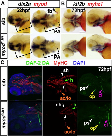
Early cranial skeletal development is normal in myodfh261 mutants. A,B. In situ mRNA hybridisation for dlx2a and myod (A), klf2b and myhz1 (B) in 52 hpf (A) and 72 hpf (B) myodfh261 mutant and sibling embryos, shown in wholemount, anterior to left. Outer panels, lateral viewand central panels, ventral view. Myod mRNA is absent but dlx2a unaffected in the pharyngeal arches (PA, brackets). Myhz1 mRNA is detected in sternohyoideus (sh, arrows) but not in other head muscles in the mutant. klf2b mRNA in skeletal parts is normal. C. Confocal stacks of heads of myodfh261 mutant and sibling stained with DAF-2DA (green), MyHC (MF20, red) and DAPI (blue). Anterior to left, ventrolateral view. Muscle in the mutant is missing except for the sternohyoideus (sh, white arrows), adductor operculi and levator operculi (ao and lo, orange arrows). Bones such as the opercle (op, yellow arrowhead), cleithrum (cl, pink arrowheads) and parasphenoid (ps, white arrowheads) are normal at this stage. Bar = 100 μm. |
Reprinted from Developmental Biology, 358(1), Hinits, Y., Williams, V.C., Sweetman, D., Donn, T.M., Ma, T.P., Moens, C.B., and Hughes, S.M., Defective cranial skeletal development, larval lethality and haploinsufficiency in Myod mutant zebrafish, 102-12, Copyright (2011) with permission from Elsevier. Full text @ Dev. Biol.