
FIGURE
Fig. 1
- ID
- ZDB-FIG-111018-39
- Publication
- Yu et al., 2010 - Evolving Cardiac Conduction Phenotypes in Developing Zebrafish Larvae: Implications to Drug Sensitivity
- Other Figures
- All Figure Page
- Back to All Figure Page
Fig. 1
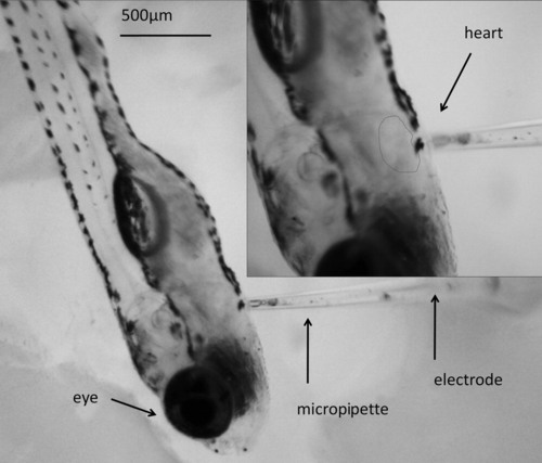
Positioning micropipette-based electrodes to a zebrafish larva. The recording micropipette suction electrode was positioned in close contact with ventral part of the larvae above the contracting hearts (noted in dashed line). A slight negative pressure was applied to secure contact between the micropipettes and larvae. The representative larva was recorded at 14 days postfertilization (dpf). |
Expression Data
Expression Detail
Antibody Labeling
Phenotype Data
Phenotype Detail
Acknowledgments
This image is the copyrighted work of the attributed author or publisher, and
ZFIN has permission only to display this image to its users.
Additional permissions should be obtained from the applicable author or publisher of the image.
Full text @ Zebrafish