
Fig. 2
- ID
- ZDB-FIG-110914-27
- Publication
- Shono et al., 2011 - Acquisition of glial cells missing 2 Enhancers Contributes to a Diversity of Ionocytes in Zebrafish
- Other Figures
- All Figure Page
- Back to All Figure Page
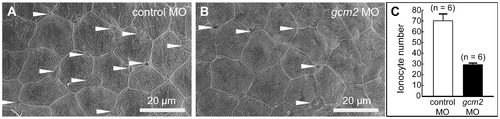
Scanning electron microscopy of the yolk sac membrane in 3-day-old zebrafish. (A, B) Magnification of the yolk sac membrane. (A) An embryo injected with a control morpholino (MO). (B) An embryo injected with a gcm2 MO. (C) Quantitative comparison of the ionocyte number per 0.04 mm2 between embryos injected with control MO and gcm2 MO. The ionocyte number on the yolk sac membrane area in gcm2 morphants is less than that in control MO. Arrowheads indicate ionocytes. Because it is likely that cells were still alive or had differentiated without expression of a marker gene by loss of function, we observed the external morphology of the skin surface in morphants using scanning electron microscopy. |
| Fish: | |
|---|---|
| Knockdown Reagent: | |
| Observed In: | |
| Stage: | Protruding-mouth |