
Fig. 2
- ID
- ZDB-FIG-110719-2
- Publication
- Tryon et al., 2011 - Lineage relationship of direct-developing melanocytes and melanocyte stem cells in the zebrafish
- Other Figures
- All Figure Page
- Back to All Figure Page
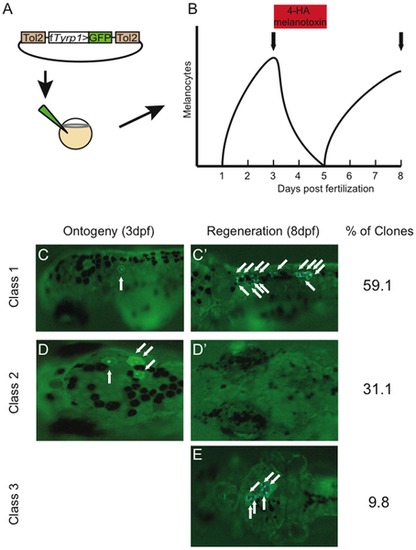
Three classes of melanocyte clones are revealed from clonal analysis. (A) Plasmid construct containing the melanocyte specific Takifugu rubripes Tyrosinase related protein 1 promoter driving GFP (fTyrp1>GFP) and flanked by Tol2 transposon arms was co-injected with transposase mRNA into 1–2 cell embryos. (B) Experimental protocol used for clonal analysis of melanocyte lineages. Following transposon injection, ontogenetic melanocytes are allowed to develop normally through 3 dpf, and then screened for GFP+ melanocytes. 4-HA treatment between 3 and 5 dpf was then used to ablate melanocytes. Drug was then washed out to allow melanocyte regeneration from MSCs. At 8 dpf fish are again screened for the presence of GFP in regeneration melanocytes. Vertical arrows indicate time of scoring for labeled melanocytes. (C–E) Representative pictures of fish showing ontogenetic melanocytes prior to ablation (C and D) and regeneration melanocytes following ablation and regeneration (C′ and E). Note that the same fish is shown in C and C′ as well as D and D′. Arrows point to GFP expressing ontogenetic and regeneration melanocytes. Fish with GFP labeled melanocytes can be divided in 3 clone classes: Class 1, with both ontogenetic and regeneration melanocytes labeled, indicating integration in a bipotent melanogenic precursor; Class 2, with only ontogenetic melanocytes labeled, indicating integration in a restricted direct-developing precursor; and Class 3, with only regeneration melanocytes labeled, indicating integration in a restricted MSC precursor. |