
Fig. 2
- ID
- ZDB-FIG-110712-67
- Publication
- Burguière et al., 2011 - Alkali-like myosin light chain-1 (myl1) is an early marker for differentiating fast muscle cells in zebrafish
- Other Figures
- All Figure Page
- Back to All Figure Page
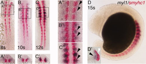
Expression of myl1 during early- to mid-somitogenesis. Expression of myl1 was detected in the paraxial mesoderm by whole mount in situ hybridization from the 8-somite stage. A: myl1 (blue) and smyhc1 (red) expression in flat-mounted and (A2) transverse section in an 8-somite-stage embryo. A3: Blow-up showing myl1 expression (arrowheads) concentrated to the posterior part within the somite and absent from the smyhc1-labelled adaxial cells. B: myl1 (blue) and smyhc1 (red) expression in flat-mounted and (B2) transverse section in a 10-somite-stage embryo. B3: Blow-up showing myl1 expression (arrowheads) concentrated to the medio-posterior part within the somite and restricted to non-adaxial cells of the paraxial mesoderm. C: myl1 (blue) and smyhc1 (red) expression in flat-mounted and (C2) transverse section in a 12-somite-stage embryo. C3: Blow-up of the myl1-expressing region (arrowheads). Myl1 (blue) is not co-expressed with smyhc (red) and its expression is concentrated to the medio-posterior part of the somites. D: myl1 expression in 15-somite-stage embryo. D2: Blow-up of a single dissected somite showing the distinct myl1- (black arrowhead) and smyhc1- (white arrowhead) expressing regions. |