
Fig. 8.
- ID
- ZDB-FIG-110712-27
- Publication
- Bruses, 2011 - N-cadherin regulates primary motor axon growth and branching during zebrafish embryonic development
- Other Figures
- All Figure Page
- Back to All Figure Page
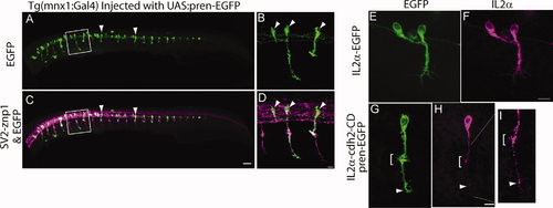
Tg(mnx1:Gal4-VP16) embryos were injected at the one-cell stage with a plasmid encoding pren-EGFP under a 14X-UAS element, fixed at 24 hpf, and immunostained with SV2 and znp1 antibodies. Arrowheads point to primary motor neurons expressing EGFP. B,D: Higher magnification images of the boxed areas in A,C, respectively. Arrowheads point to primary motor neuron somas. E–I: Tg(mnx1:Gal4-VP16) embryos were injected at the one-cell stage with the 14X-UAS-IL2α-EGFP plasmid (E,F) or with 14X-UAS-IL2α-CD and pren-EGFP plasmid (G–I). Embryos were fixed at 24 hpf and immunostained with anti-IL2α antibodies (F,H,I). IL2α-EGFP is evenly distributed throughout the motor neuron (E,F). Pren-EGFP labels the entire cell body and axon (G), whereas IL2α-CD is detected as discrete puncta through out the cell (H, arrowheads). I: Higher magnification of the axon shown in G, indicating IL2α-CD puncta accumulated at the choice point (bracket) and at the distal tip of the axon. Dorsal is to the top and rostral is to the left. Scale bars = 50 μm in C (applies to A,C); 10 µm in D (applies to B,D); 10 μm in F (applies to E,F); 10 μm in H (applies to G,H). |