
Fig. 5
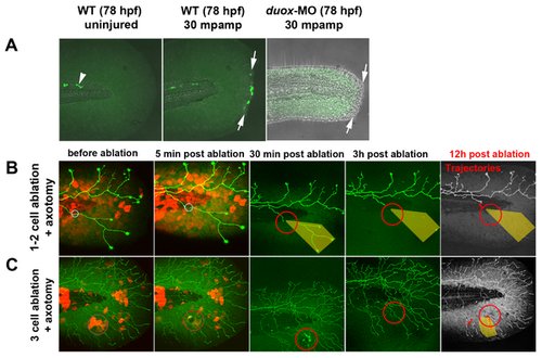
(A) In uninjured fins, H2O2 was always detectable with pentafluorobenzenesulfonyl fluorescein (see Methods) in a few cells near the notochord (arrowhead) but was not detectable in fin tissue. Fin amputation (arrows) produced high levels of H2O2 in wound marginal cells at 30 min post-amputation. In contrast, H2O2 levels were either undetectable or very low in duox1 morphants at 30 mpamp. (B, C) Ablating multiple keratinocytes produced hydrogen peroxide (H2O2). Time-lapse sequences from 78–90 hpf. The rightmost panel shows axon tip trajectories (red) over the course of the time-lapse and denervated territory (shaded areas). (B) Ablation of one or two keratinocytes (circles) did not produce sufficient H2O2 to be detected by the H2O2 sensor pentafluorobenzenesulfonyl fluorescein and did not promote axon regeneration of a nearby severed axon. (C) Ablating more (e3) keratinocytes (circles) induced H2O2 production around the wound margin, as detected with the H2O2 sensor, and improved regeneration of a nearby axotomized arbor, which grew into denervated territory (shaded area, trajectories). mpamp, minutes post amputation. |