
Fig. S1
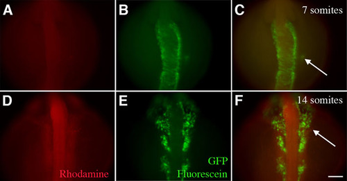
Arterial pole progenitors are not derived from neural crest. Ventral view, with cranial to top, of zebrafish after photoactivation of cells at the 7 somite stage. (A-C) When cells in grid zone F were labeled using caged fluorescein:rhodamine dextran (1:1 v/v) in Tg(sox10::eGFP) zebrafish at the 7 somite stage, the labeled cells (green, red and arrow) were clearly distinct from the GFP-positive neural crest cells in the neural tube. (D-F) By the 14-somite stage, the labeled cells (arrow) began to converge with the migrating neural crest but were still not overlapping. The GFP-positive neural crest cells were migrating from the neural tube at this time. Scale bar: 10 μm. |