
FIGURE
Fig. 4
- ID
- ZDB-FIG-110405-14
- Publication
- Choi et al., 2011 - Aplexone targets the HMG-CoA reductase pathway and differentially regulates arteriovenous angiogenesis
- Other Figures
- All Figure Page
- Back to All Figure Page
Fig. 4
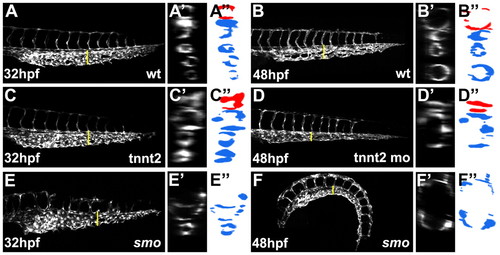
Blood circulation is not required for sprouting angiogenesis from the caudal vein. (A,B) Caudal vein plexus in wild-type Tg(kdrl:GFP) embryos observed at 32 hpf (A) and 48 hpf (B). (C-F) At 32 hpf, the primordial caudal vein plexus was properly formed in tnnt2 morphant (C) and smo mutant (E) embryos, but the plexus structure was not maintained and degenerated into a single-lumen structure at 48 hpf (D,F). (A′-F′) Optical cross-sections of blood vessels indicated by yellow lines in A-F. (A"-F") Schematic drawings of images shown in A′-F′. Red and blue represent the artery and vein, respectively. |
Expression Data
Expression Detail
Antibody Labeling
Phenotype Data
Phenotype Detail
Acknowledgments
This image is the copyrighted work of the attributed author or publisher, and
ZFIN has permission only to display this image to its users.
Additional permissions should be obtained from the applicable author or publisher of the image.
Full text @ Development