
FIGURE
Fig. s2
- ID
- ZDB-FIG-110328-2
- Publication
- Chun et al., 2011 - Fli+ etsrp+ Hemato-Vascular Progenitor Cells Proliferate at the Lateral Plate Mesoderm during Vasculogenesis in Zebrafish
- Other Figures
- All Figure Page
- Back to All Figure Page
Fig. s2
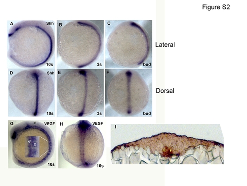
A-F are whole mount Shh ISH embryos at 10 som (A, D), 3 som (B, E) and bud (C, F) embryonic stage with expression noticed at midline in all stages. (A-C) is lateral view and, (D-F) is dorsal view. (G-H) are whole mount vegf ISH at 10 som. Inset in G shows the vegf expression in endoderm (e) adjoining the yolk and somite (s). I is immunostaining of 10 som section for VEGF protein. Yellow arrow indicates hypochord staining of VEGF protein. |
Expression Data
Expression Detail
Antibody Labeling
Phenotype Data
Phenotype Detail
Acknowledgments
This image is the copyrighted work of the attributed author or publisher, and
ZFIN has permission only to display this image to its users.
Additional permissions should be obtained from the applicable author or publisher of the image.
Full text @ PLoS One