
Fig. 4
- ID
- ZDB-FIG-110317-31
- Publication
- Yoo et al., 2010 - Differential Regulation of Protrusion and Polarity by PI(3)K during Neutrophil Motility in Live Zebrafish
- Other Figures
- All Figure Page
- Back to All Figure Page
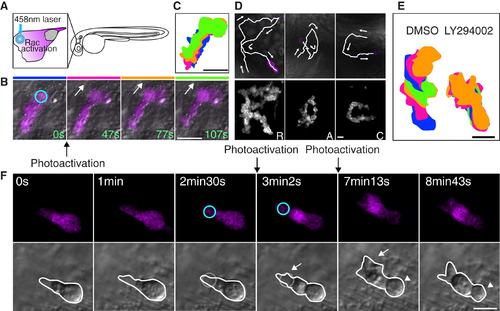
Photoactivation of Rac at the Leading Edge Can Rescue the Protrusion Defects but Not the Rounded Tail or Migration Defects Induced by PI(3)K Inhibition (A) A schematic representation of photoactivation of Rac at the neutrophil leading edge in zebrafish. (B) Photoactivation of Rac at the leading edge induces protrusion and migration of a neutrophil in tissues (Movie S5). The circle indicates the position of Rac photoactivation. Scale bar = 20 μm. (C) Overlayed images of (B) show directional migration induced by photoactivated Rac. Scale bar = 20 μm. (D) Spelling by neutrophil trajectories guided through repetitive photoactivation of Rac at the leading edge (Movie S6). Scale bars in (D–F) = 10 μm. (E) Overlayed images show that PI(3)K inhibition disturbs Rac photoactivation-induced migration. (The leading edge was activated for 20 s twice during 5 min imaging). (F) Photoactivation of Rac at the front (circles) can rescue the protrusion defect induced by PI(3)K inhibition (arrows), but not the rounded tail defect (arrowheads) (Movie S8). Images are representative of more than five time-lapse movies from experiments repeated on at least two separate dates. |
Reprinted from Developmental Cell, 18(2), Yoo, S.K., Deng, Q., Cavnar, P.J., Wu, Y.I., Hahn, K.M., and Huttenlocher, A., Differential Regulation of Protrusion and Polarity by PI(3)K during Neutrophil Motility in Live Zebrafish, 226-236, Copyright (2010) with permission from Elsevier. Full text @ Dev. Cell