
Fig. S3
- ID
- ZDB-FIG-110128-42
- Publication
- d'Alencon et al., 2010 - A high-throughput chemically induced inflammation assay in zebrafish
- Other Figures
- All Figure Page
- Back to All Figure Page
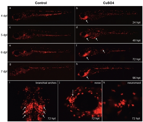
Behavior of leukocytes after long-term copper exposure in zebrafish larvae. At 3 days postfertilization (dpf), transgenic lysC::DsRED2 fish were left untreated (a, c, e, and g) or exposed permanently thereafter to 10 μM CuSO4 (b, d, f, h, and i-k) and were imaged daily until 7 dpf (times in the right-hand column are expressed in hours posttreatment, hpt). (a-h) Lateral views of entire larvae. Note the general dispersal of leukocytes in treated vs. control fish simultaneous with accumulation in different anterior regions, especially the branchial arches of the animal beginning 1 day after beginning treatment. (i-k) Closeups of specific areas at 72 hpt. (i) Ventral view of branchial arches. (j) Lateral view of head; arrow indicates olfactory pit area. Closeup view of the area surrounding a neuromast. Note that fish exposed for long periods to copper sulfate suffer developmental delays. |