
FIGURE
Fig. 4
- ID
- ZDB-FIG-101124-20
- Publication
- Tschopp et al., 2010 - Funduscopy in adult zebrafish and its application to isolate mutant strains with ocular defects
- Other Figures
- All Figure Page
- Back to All Figure Page
Fig. 4
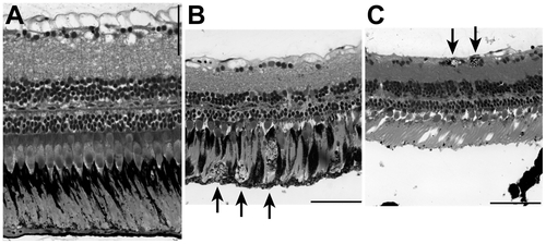
Adult retina sections. (A) Wild type zebrafish. (B) Right eye and (C) left eye of a fish with black spots in the retina; the funduscopy of this fish is shown in Figure 3 (H). Arrows indicate cells with black granules: likely macrophages filled with RPE cells or detached RPE cells. The detached RPE in (C) is a histological artefact. Scale bar: 50 μm. |
Expression Data
Expression Detail
Antibody Labeling
Phenotype Data
| Fish: | |
|---|---|
| Observed In: | |
| Stage: | Adult |
Phenotype Detail
Acknowledgments
This image is the copyrighted work of the attributed author or publisher, and
ZFIN has permission only to display this image to its users.
Additional permissions should be obtained from the applicable author or publisher of the image.
Full text @ PLoS One