
Fig. S2
- ID
- ZDB-FIG-101118-49
- Publication
- Hesselson et al., 2009 - Distinct populations of quiescent and proliferative pancreatic β-cells identified by HOTcre mediated labeling
- Other Figures
- All Figure Page
- Back to All Figure Page
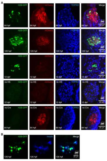
HOTcre controls. (A and B) Confocal sections of islets stained with TOPRO (A) to mark nuclei. (A) The mCherry induction reporter was induced ubiquitously and H2B-GFP was expressed specifically in β-cells by heat-shock of Insulin-HOTcre embryos at 24 hpf. By 84 hpf, mCherry is expressed at low levels in many tissues. Within the slowly dividing endocrine compartment, mCherry is initially retained as bright puncta. The intensity of the mCherry signal is reduced by 120 hpf and reaches the limit of detection by 12 dpf. Without heat-shock treatment, H2B-GFP is not detected in the islet. In the absence of Tg(ins:Cre; cryaa:Venus)s924, H2B-GFP is not detected in the islet following heat-shock treatment at 24 hpf. (B) H2B-GFP was expressed specifically in β-cells by heat-shock of Insulin-HOTcre embryos at 120 hpf. By 126 hpf, H2B-GFP is expressed in all Insulin positive cells. |