
Fig. S7
- ID
- ZDB-FIG-101108-36
- Publication
- Jänicke et al., 2010 - Zebrafish grainyhead-like1 is a common marker of different non-keratinocyte epidermal cell lineages, which segregate from each other in a Foxi3-dependent manner
- Other Figures
- All Figure Page
- Back to All Figure Page
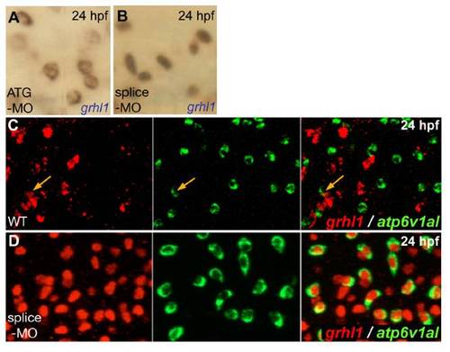
grhl1 morphants display grhl1 expression in all ionocytes, with nuclear localisation when grhl1-splice MO, and cytoplasmic localisation when grhl1-ATG MO is used. All panels show in situ hybridisations at 24 hpf and with probes indicated in lower right corners. (A,B) Grhl1 transcripts were located in the nucleus upon injection of grhl1-splice-MO (B; also used in D), whereas they were cytoplasmic when grhl1-ATG-MO was used (A). (C,D) Double fluorescent in situ hybridisations indicating that in wild-type embryos (WT), only few ATPase6v1al-positive ionocytes (in green) were grhl1-positive (in red) (C; orange arrows), whereas in the grhl1 morphant (D), all ATPase6v1al-positive ionocytes contained nuclear grhl1 transcripts; anterior trunk region; left and middle pictures show single channels, right picture merged image. |