|
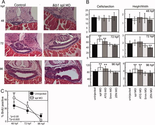
Knockdown of lkb1 stimulates epithelial cell proliferation and growth. A: Histological sections through the intestine of uninjected, spl morphant, and ATG morphant, embryos at 48, 72, and 96 hpf. At 48 hpf, the gut lumen is outlined by solid black line. g, gut. Shown are representative sections that correspond to the median gut size as determined by morphometry and depicted in the bar graph. Sections were stained with H&E and original magnification was 400x. B: Depiction of morphometric analysis for cell number and cell shape (height-to-width ratio) using 25 embryos per treatment. A-P positions were matched between embryos using standard anatomic landmarks (pancreatic islet, gall bladder, neural tube). C: Analysis of proliferation rate by BrdU incorporation comparing uninjected (black squares) and spl morphant (white squares) at 48, 72, and 96 hpf. Significance was determined using Student′s two-tail, unpaired t-test (n=25 embryos). *P < 0.05; **P < 0.005.
|