
Fig. 10
- ID
- ZDB-FIG-101025-7
- Publication
- Larson et al., 2010 - Defective adult oligodendrocyte and Schwann cell development, pigment pattern, and craniofacial morphology in puma mutant zebrafish having an alpha tubulin mutation
- Other Figures
- All Figure Page
- Back to All Figure Page
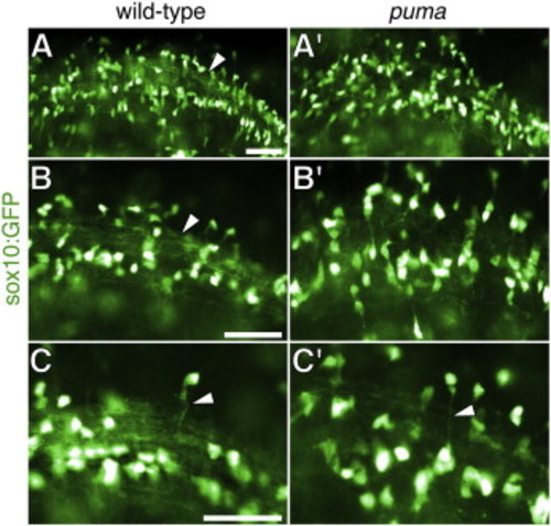
Defects in oligodendrocyte patterning and myelination in puma mutant early larvae. (A–C) sox10:GFP transgene reveals oligodendrocyte cell bodies and processes, that are more disorganized in puma mutants than in wild-type at 5 dpf. Cell bodies are deliberately overexposed (shown here in white) to reveal fainter processes. (A, A′) Low magnification showing the orderly array of myelinating processes in wild-type (arrow), and more disorganized processes in puma mutants. (B, B′) Higher magnification of different individuals. (C, C′) Relatively long processes are observed in both wild-type and puma mutants (arrowheads). Scale bars: in A, 40 μm for A, A′; in 40 μm for B, B′; in C, 40 μm for C, C′. |
| Gene: | |
|---|---|
| Fish: | |
| Anatomical Terms: | |
| Stage: | Day 5 |
| Fish: | |
|---|---|
| Observed In: | |
| Stage: | Day 5 |
Reprinted from Developmental Biology, 346(2), Larson, T.A., Gordon, T.N., Lau, H.E., and Parichy, D.M., Defective adult oligodendrocyte and Schwann cell development, pigment pattern, and craniofacial morphology in puma mutant zebrafish having an alpha tubulin mutation, 296-309, Copyright (2010) with permission from Elsevier. Full text @ Dev. Biol.