
FIGURE
Fig. 7
- ID
- ZDB-FIG-101021-36
- Publication
- Lyons et al., 2010 - Carboxypeptidase A6 in zebrafish development and implications for VIth cranial nerve pathfinding
- Other Figures
- All Figure Page
- Back to All Figure Page
Fig. 7
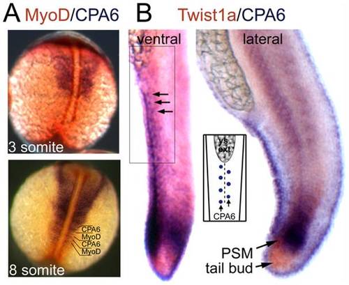
Distribution of CPA6 mRNA compared with somitogenesis markers. In situ hybridization was performed with RNA probes specific for (A) CPA6 (purple) and MyoD (orange) at 3 and 8 somite stages (11 and 14 hpf), and (B) CPA6 (purple) and Twist1b (orange) at 24 hpf. Arrows indicate ectodermal cells arranged along the ventral ridge of the tail and expressing CPA6. The regular arrangement of these cells, also found along the dorsal ridge, is illustrated in the inset. PSM, presomitic mesoderm; ys ext, yolk-sac extension. |
Expression Data
| Genes: | |
|---|---|
| Fish: | |
| Anatomical Terms: | |
| Stage Range: | 1-4 somites to Prim-5 |
Expression Detail
Antibody Labeling
Phenotype Data
Phenotype Detail
Acknowledgments
This image is the copyrighted work of the attributed author or publisher, and
ZFIN has permission only to display this image to its users.
Additional permissions should be obtained from the applicable author or publisher of the image.
Full text @ PLoS One