|
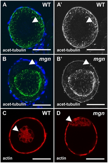
Localization of stable microtubules but not actin is disrupted in mgn mutant oocytes. Stage I oocytes stained with an antibody to acetylated tubulin to label stable microtubules (A,B) or rhodamine phalloidin to label actin cytoskeleton (C,D). In wild-type stage I oocytes, acetylated microtubules are uniformly distributed throughout the oocyte (A,A′), whereas in mgn mutant oocytes, acetylated microtubules are largely absent from peripheral regions of the oocyte (B,B′). DAPI staining around oocytes labels nuclei of surrounding somatic follicle cells. Green = acetyated tubulin; blue = DAPI. In both wild-type (C) and mgn mutant oocytes (D), actin is localized to the nucleus and at the cortex. Arrowheads indicate oocyte nuclei. Red = phalloidin. Scale bars = 25 microns. All images are single optical sections.
|