|
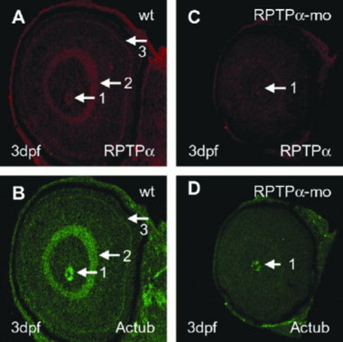
Defects in retinal organization and differentiation in receptor protein-tyrosine phosphatase alpha morpholino (RPTPα-mo) -injected embryos. A,B: Oblique section of 3-days-postfertilization (dpf) -old wild-type (wt) embryo labeled with polyclonal anti-RPTPα antibody (AP5478; A) and anti-acetylated tubulin (B). Labeling is found in the optic nerve (arrow 1), the inner plexiform layer (arrow 2), and the outer plexiform layer (arrow 3). C,D: Oblique section of 3-dpf-old RPTPα-mo–injected embryo labeled with polyclonal anti-RPTPα antibody (AP5478; C), and anti-acetylated tubulin (Actub) antibody (D). Labeling of RPTPα and acetylated tubulin is found in the optic nerve (arrow 1), all other structures labeled in wild-type are not labeled in RPTPα-mo–injected embryos.
|