
FIGURE
Fig. S3
- ID
- ZDB-FIG-100706-28
- Publication
- Sato et al., 2010 - Single-cell analysis of somatotopic map formation in the zebrafish lateral line system
- Other Figures
- All Figure Page
- Back to All Figure Page
Fig. S3
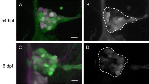
Distribution of newly differentiated neurons in the posterior lateral line (PLL) ganglion. A,C: The neurons labeled magenta has been differentiated and photoconverted at 30 hours postfertilization (hpf; A) and 5 days postfertilization (dpf; C). The cells expressing only green Kaede are newly added neurons within 24 hr just before the observation. B,D: Z-stack images of newly added neurons abstracted from the raw images (A, C) by image processing. The outline of PLL ganglion is indicated by a white dotted line. Scale bars = 10 μm. |
Expression Data
Expression Detail
Antibody Labeling
Phenotype Data
Phenotype Detail
Acknowledgments
This image is the copyrighted work of the attributed author or publisher, and
ZFIN has permission only to display this image to its users.
Additional permissions should be obtained from the applicable author or publisher of the image.
Full text @ Dev. Dyn.