Fig. 6
- ID
- ZDB-FIG-100525-35
- Publication
- Okuda et al., 2010 - B1 SOX coordinate cell specification with patterning and morphogenesis in the early zebrafish embryo
- Other Figures
- All Figure Page
- Back to All Figure Page
|
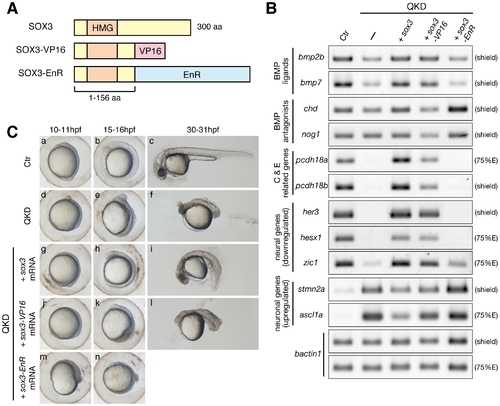
Regulatory actions of the B1 SOX proteins. (A) Schematic representation of the protein structures of SOX3, SOX3-VP16 and SOX3-EnR. Dominant activator and repressor forms of SOX3 were constructed by fusing the VP16 activation and Engrailed repression (EnR) domains, respectively, to truncated SOX3(1–156 aa). (B) Gene expression responses to SOX3, SOX3-VP16 or SOX3-EnR under QKD conditions. mRNAs of sox3, sox3-VP16 and sox3-EnR (20 pg) were individually injected with the MOs for QKD and gene expression responses were examined by RT-PCR. The exogenous supply of either SOX3 or SOX3-VP16 but not by SOX3-EnR recovered expression of genes that were downregulated (bmp2b/7, pcdh18a/18b, her3, hesx1 and zic1) in the QKD embryos and also suppressed expression of genes that were upregulated (stmn2a and ascl1a). bactin1 was used as an RT-PCR control. (C) Partial rescue by SOX3-VP16 and strengthening by SOX3-EnR of the morphological phenotypes of the QKD embryos. Live embryo images at 10–11, 15–16, and 30–31 hpf were observed. SOX3-VP16-injected embryos showed a rather ventralized phenotype. Embryos coinjected with SOX3-EnR died during late segmentation stages. All are lateral views. |

