
Fig. 2
- ID
- ZDB-FIG-100322-13
- Publication
- Kimmel et al., 2010 - Modes of developmental outgrowth and shaping of a craniofacial bone in zebrafish
- Other Figures
- All Figure Page
- Back to All Figure Page
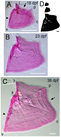
Time-course of shape changes and growth of the opercle in the late larva and in young adult fish. (A) 18 dpf. (B) 23 dpf. (C) 38 dpf. (D) Silhouettes scaled to the same final magnification to illustrate the amount of overall bone growth during this period. Imaging used oblique incident lighting to reveal matrix morphology. Incremental bands, the joint socket, and thickened, relatively heavily mineralized struts at characteristic positions along the bone are particularly well shown in C. The refractile fibers at the j apex in C are remains of the tendon that attached the dilator operculi muscle to the bone at this apex. Note a concave curvature along the vp edge (arrow in C) that was not present during the larval stages shown in Figure 1. Abbreviations and orientations as in Figure 1. Scale bars: 200 μm. |