FIGURE

Fig. 5

ID
ZDB-FIG-100319-20
Publication
Ko et al., 2010 - Broad-Minded Links Cell Cycle-Related Kinase to Cilia Assembly and Hedgehog Signal Transduction
Other Figures
All Figure Page
Back to All Figure Page
Fig. 5

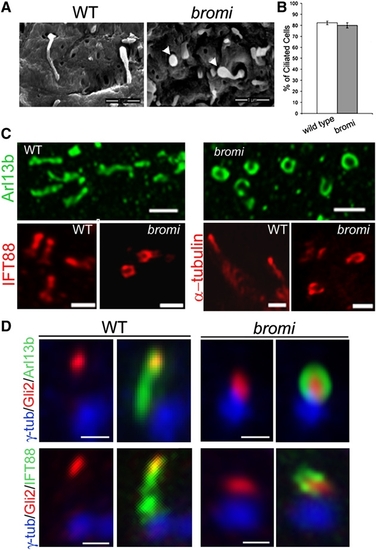
Cilia Defects in bromi Mutants
(A) Neuroepithelial cilia from E10.5 WT and bromi mutant neural tubes analyzed by scanning electron microscopy. bromi mutant cilia showed a swollen or bulbous morphology (arrowheads). Scale bars, 1 μm.
(B) Cilia frequency in the WT and bromi mutant neuroepithelium. Error bars represent ± 1 SEM.
(C) Confocal images of neural tube cilia stained with Arl13b (upper panels), IFT88 and acetylated α-tubulin (lower panels). Scale bars, 1 μm.
(D) Neural tube cilia from E10.5 WT and bromi mutant embryos immunostained for γ-tubulin/Arl13b/ Gli2 or γ-tubulin/IFT88/Gli2. Scale bars, 0.5 μm.

Expression Data

Expression Detail
Antibody Labeling
Phenotype Data

Phenotype Detail
Acknowledgments
This image is the copyrighted work of the attributed author or publisher, and ZFIN has permission only to display this image to its users. Additional permissions should be obtained from the applicable author or publisher of the image.

Reprinted from Developmental Cell, 18(2), Ko, H.W., Norman, R.X., Tran, J., Fuller, K.P., Fukuda, M., and Eggenschwiler, J.T., Broad-Minded Links Cell Cycle-Related Kinase to Cilia Assembly and Hedgehog Signal Transduction, 237-247, Copyright (2010) with permission from Elsevier. Full text @ Dev. Cell