
Fig. 5
- ID
- ZDB-FIG-100302-37
- Publication
- Loynes et al., 2010 - Pivotal Advance: Pharmacological manipulation of inflammation resolution during spontaneously resolving tissue neutrophilia in the zebrafish
- Other Figures
- All Figure Page
- Back to All Figure Page
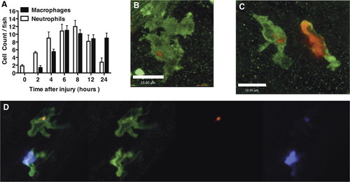
Macrophages take up apoptotic neutrophils during inflammation resolution in vivo. (A) Three dpf mpx:GFP zebrafish were injured as described previously. Two hours before the time indicated, larvae were stained with neutral red for 2 h. The number of neutral red-positive macrophages and GFP-positive neutrophils was counted for each fish under bright-field illumination and 488 nm fluorescent illumination, respectively. (B and C) Seven dpf wild-type zebrafish were injured and fixed 12 h later in 4% paraformaldehyde at 4°C, followed by staining for mpx activity and L-plastin immunohistochemistry, as described in Materials and Methods. Single macrophages can be seen containing TSA-positive neutrophilic material. (C) A neutrophil is shown alongside for comparison. (D) Cy5-TSA staining (purple) was combined with L-plastin staining (green) and TUNEL staining (red) to show colocalization of apoptotic markers and neutrophil markers within a macrophage. Three-dimensional reconstructions are shown in Supplemental Movies 4 and 5 to confirm location of the Cy-3 TSA-positive material completely within the macrophage. Images were taken using a x60 Plan Apo Oil immersion NA1.40 (Olympus). |