
Fig. 8
- ID
- ZDB-FIG-091221-38
- Publication
- Wiles et al., 2009 - Use of Zebrafish to Probe the Divergent Virulence Potentials and Toxin Requirements of Extraintestinal Pathogenic Escherichia coli
- Other Figures
- All Figure Page
- Back to All Figure Page
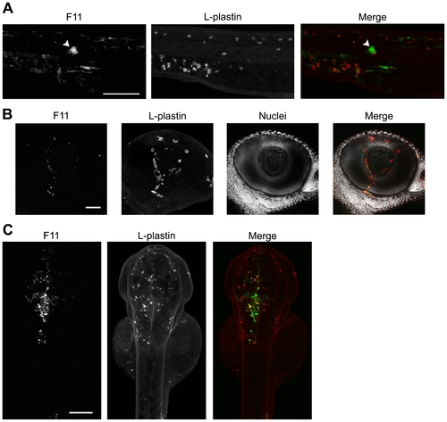
ExPEC isolates capable of blood colonization can disseminate and persist in a variety of host microenvironments. F11, carrying pGEN-GFP(LVA) for constitutive expression of destabilized GFP (green), was inoculated into the blood using a dose of 4,000–6,500 CFU. Samples were fixed at 6 or 12 hpi and phagocytes (red) were labeled using L-plastin-specific antibody for visualization by fluorescent confocal microscopy. (A) 20X z-projection of the tail region from an F11-infected embryo at 12 hpi. The arrowhead indicates a commonly observed bacterial microcluster. (B) 40X z-projection of the eye from an embryo at 6 hpi with F11. Hoechst nuclear dye (grey) was used to highlight the anatomical structure of the eye. (C) Dorsal to ventral 10X z-projection of the head region of an embryo at 12 hpi with F11. Scale bars = 100 μm for (A) and (C) and 50 μm for (B). |