
Fig. 1
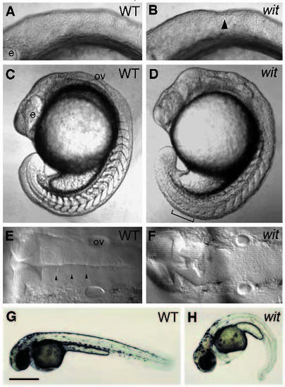
Phenotype of live witta52b embryos. If not specified, all the following figures are rostral to the left, dorsal to the top. Lateral views of wild-type (A) and mutant (B) embryos at 9-somite stage. The indentation in the hindbrain (arrowhead) is indicated. Lateral views of wild-type (C) and mutant (D) embryos at 17-somite stage. The indistinct somite boundaries (bracket) are indicated. Dorsal views of wild-type (E) and mutant (F) embryos at 26 hours postfertilization (hpf). Normal rhombomeric boundaries (arrowheads) are not seen in mutant embryos. Lateral views of wildtype (G) and mutant (H) embryos at 36 hpf. Note that there are no visible pigmented melanophores in the posterior trunk of mutant embryo in H. e, eye; ov, otic vesicle. Bar, 200 µm (A,B,E,F); 335 µm (C,D); 640 µm (G,H). |
| Fish: | |
|---|---|
| Observed In: | |
| Stage Range: | 5-9 somites to Prim-25 |