|
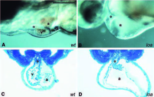
Chamber demarcation mutant. Images of the hearts of live 2-day old wild-type (A) and loatu29d (B) embryos are shown. The atrium (a) is significantly enlarged in loatu29d. No obvious structure representing the ventricle (v) is seen between the atrium and the outflow tract (o). The atrium, ventricle and outflow tract are present in a transverse section of the heart in a 2-day old wild-type embryo (C). However, no obvious structure representing the ventricle is seen in a transverse section of the heart of the 2-day old loatu29d embryo (D). The embryos are oriented with the anterior to the left and dorsal to the top. Scale bar, 100 μm.
|