
FIGURE
Fig. 2
Fig. 2
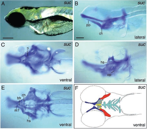
Photomicrographs of lateral views (A,B,D) and ventral views (C,E,F) of sucker mutant embryos reveal that Meckel’s cartilage (lower jaw) is strongly reduced or absent. The diagram (F) represents a composite of elements photographed in C and E. The neurocranium is omitted. The ceratohyal is reduced and fused to the basihyal (E). Although the posterior arches are present, they are reduced. Ventral to the ceratohyal additional unidentified plate-like cartilaginous elements can be detected (C,F). Scale bars: 200 μm (A); 100 μm (B-F). |
Expression Data
Expression Detail
Antibody Labeling
Phenotype Data
| Fish: | |
|---|---|
| Observed In: | |
| Stage Range: | Protruding-mouth to Day 5 |
Phenotype Detail
Acknowledgments
This image is the copyrighted work of the attributed author or publisher, and
ZFIN has permission only to display this image to its users.
Additional permissions should be obtained from the applicable author or publisher of the image.
Full text @ Development