
Fig. 1
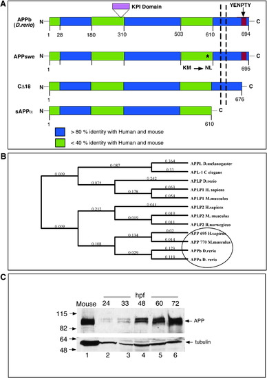
Homology and phylogenetic analysis of APP. (A) Schematic representation of zebrafish APPb and the truncation mutants of human APP used in this study. Zebrafish APPb is a 694-residue-long protein with the extracellular N-terminal domain, a transmembrane domain (shown by the vertical dashed lines), and an intracellular C-terminal domain that contains the EYNPTY motif (red box). APP family proteins show 3 highly conserved domains (blue regions) that are more than 80% identical among each other, and are separated by less conserved regions (green regions) that are less than 40% identical at the protein level. APPa contains a 40-residue-long KPI domain (shown in purple), which is not present in APPb. The location of the Swedish mutation in APP (where residues ‘KM’ are mutated to ‘NL’) is shown by an ∗. APP-CΔ18 lacks the last 18 C-terminal residues, including the EYNPTY motif. Soluble sAPPα, which is truncated at the α-cleavage site and lacks both the membrane-spanning domain and the intracellular domain. The percentage homology is obtained by using Blast at ncbi.nlm.nih.gov or ClustalW in MacVector 7.2.2, both using Blosum matrices. (B) Phylogenetic groupings between APP family proteins from different species. The tree was generated by using default settings following multiple alignment using ClustalW and Neighbor-joining algorithms. Zebrafish APPa and APPb group closer with mouse and human APP compared to invertebrate APP proteins. (C) Zebrafish APP levels increase during embryogenesis. Western blot analysis of zebrafish proteins at different stages of development. Deyolked embryos were collected at the indicated hpf, resolved by SDS-PAGE, and probed with anti-C-terminal antibody that was raised to the last 15 residues of human APP. Total protein homogenate from a mouse brain was loaded in lane 1 as a positive control. Like mouse APP, zebrafish APP migrates as a closely spaced doublet at ∼ 100–110 kDa. The lower part of the nitrocellulose membrane was probed with anti-α-tubulin antibody (DM1A). |
Reprinted from Developmental Biology, 335(1), Joshi, P., Liang, J.O., Dimonte, K., Sullivan, J., and Pimplikar, S.W., Amyloid precursor protein is required for convergent-extension movements during Zebrafish development, 1-11, Copyright (2009) with permission from Elsevier. Full text @ Dev. Biol.