
Fig. S2
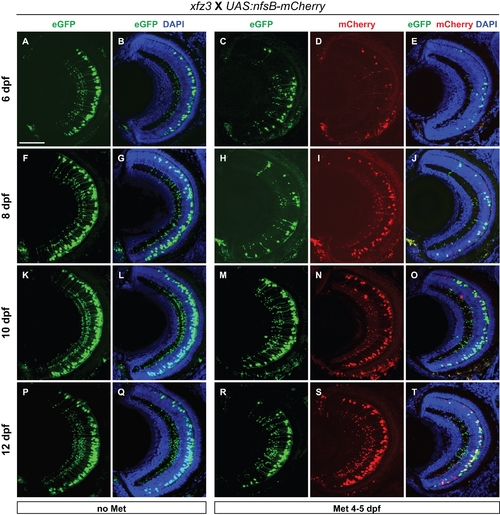
Retina recovery in larval progeny from the xfz3 × UAS:nfsB-mCherry mating. Retinal cryosections were obtained from larvae at different time points during a period of seven days following removal of Met at 5 dpf. The left panel (labelled ′no Met′ at the top) show confocal images of retina cross-sections from untreated siblings expressing only Gal4-VP16/eGFP. The right panel (labelled ′Met 4–5 dpf′) show confocal images of retina cross-sections from NTR-mCherry expressing larvae following removal of Met. The different time points and corresponding larval stages are: (C-E) 1 day post-treatment (6 dpf); (H-J) 3 days (8 dpf); (M-O) 5 days (10 dpf); (R-T) 7 days (12 dpf). The different types of fluorescence are indicated at the top. Scale bar: 50 μm. |