
Fig. 7
- ID
- ZDB-FIG-090814-24
- Publication
- Viktorin et al., 2009 - Emx3 is required for the differentiation of dorsal telencephalic neurons
- Other Figures
- All Figure Page
- Back to All Figure Page
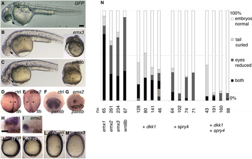
A-N: Overexpression of emx1, emx2, or emx3 mRNA, as well as emx3-homeodomain-en mRNA, dorsalizes and posteriorizes embryos. A-I,J,M: Injection of emx2 mRNA (E,G,I), emx3 mRNA (B,M), and wnt8b mRNA (C) produces embryos with posteriorized central nervous systems (loss of eyes in B,C,I loss of pax6b expression and anterior shift of egr2b expression in G; loss of otx2 expression except for an abnormally anterior expression domain in I), and dorsalization phenotypes (curly tail in B,C, insets, broadened flh-expressing notochord in E, abnormally egg-shaped embryos in E,M). J-M: Dorsalization effects are mimicked by emx3hd-en (L), but not emx3hd-VP16 (K) fusion proteins, indicating that emx3 might act as a repressor. N: Quantification of phenotypes caused by emx and wnt8b mRNA injection and their rescue by coinjection of the Wnt/beta-catenin pathway inhibitor dkk1 and Fgf pathway inhibitor spry4, suggesting that excessive emx mRNA in the early embryo potentiates the effects of these signaling pathways. A-C,H-M: Side views, dorsal to the top, rostral to the left; (D,E) Dorsal views, animal pole to the top; (F,G) Animal pole views, dorsal to the bottom. (A-C) 35 hr; (H,I) 30 hr; (D-G, J-M) bud stage. Scale bars = 100 μm. |