
Fig. 6
- ID
- ZDB-FIG-090710-67
- Publication
- Yonkers et al., 2009 - Molecular components underlying nongenomic thyroid hormone signaling in embryonic zebrafish neurons
- Other Figures
- All Figure Page
- Back to All Figure Page
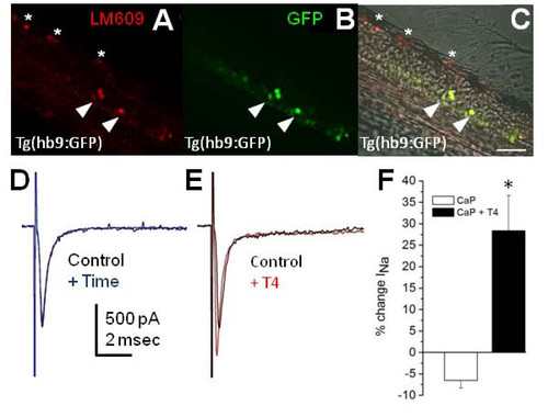
T4 increases sodium current in caudal primary motoneurons. (A-C) Tg(hb9:GFP) transgenic embryos were incubated with the αVβ3 antibody LM609. The 48-hpf Tg(hb9:GFP) transgenics displayed LM609 immunoreactivity in dorsal (asterisks) and ventral cells (arrowheads). The ventral immunoreactivity for LM609 colocalized with green fluorescent protein (GFP) in primary motoneurons (C, arrowheads). Images are oriented with dorsal neurons and ventral neurons in the upper left and lower right corners, respectively. Scale bar: 50 μm. (D, E) Caudal primary motoneuron (CaP) INa was recorded for 5 minutes (+ Time) either in the absence (D) or presence of T4 (E). Each trace shows current in response to a -10 mV depolarizing stimulus. (F) At 50 to 55 hpf, zebrafish CaPs showed rapid increases in INa amplitude in response to acute application of T4. The 30 nM T4 application significantly increased CaP INa (n = 5; P < 0.01) compared to controls (n = 5). |