
FIGURE
Fig. 6
- ID
- ZDB-FIG-090617-27
- Publication
- Nagy et al., 2009 - Endothelial cells promote migration and proliferation of enteric neural crest cells via beta1 integrin signaling
- Other Figures
- All Figure Page
- Back to All Figure Page
Fig. 6
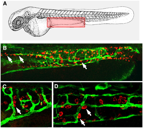
Enteric neural crest cells align with blood vessels in the zebrafish gut. (A) Diagram of a zebrafish embryo with red box depicting the approximate area of the gut imaged in panels B–D. (B) Hu+ ENCCs (red) are seen in close proximity to EGF-labeled ECs (green) in 6 dpf TG(fli1:EGFP) wild-type embryos (white arrows). This colocalization is present in both mid- (C) and posterior intestine (D). |
Expression Data
Expression Detail
Antibody Labeling
Phenotype Data
Phenotype Detail
Acknowledgments
This image is the copyrighted work of the attributed author or publisher, and
ZFIN has permission only to display this image to its users.
Additional permissions should be obtained from the applicable author or publisher of the image.
Reprinted from Developmental Biology, 330(2), Nagy, N., Mwizerwa, O., Yaniv, K., Carmel, L., Pieretti-Vanmarcke, R., Weinstein, B.M., and Goldstein, A.M., Endothelial cells promote migration and proliferation of enteric neural crest cells via beta1 integrin signaling, 263-272, Copyright (2009) with permission from Elsevier. Full text @ Dev. Biol.