|
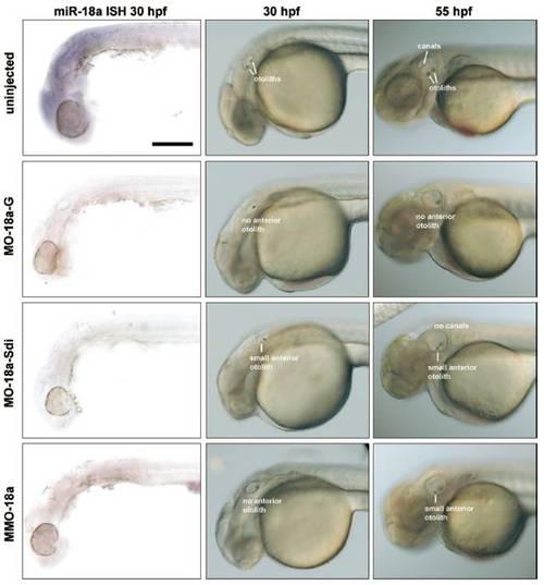
Morpholino knockdown of miR-18a using different MOs. (Left) Thirty-hpf embryos probed with miR-18a LNA probe to detect mature miRNA. (Scale bar, 200μm.) (Middle) Nomarski images of embryos at 30 hpf show either absent or smaller anterior otoliths in 18a morphants. MO-18a-Sdi morphants are either normal (data not shown) or have smaller anterior otoliths; they typically do not have a specific loss of the anterior otoliths. (Right) Nomarski images of embryos at 55 hpf showing that the phenotype of the anterior otoliths persists, although in most morphants the canals form normally. An exception are the small fraction of MO-miR-18a-Sdi morphants that continue to show reductions in anterior otolith size, which is correlated with a more general loss of canal pillar outgrowth as shown.
|