FIGURE

Fig. 3

ID
ZDB-FIG-090602-26
Publication
Warga et al., 2009 - Fate mapping embryonic blood in zebrafish: multi- and unipotential lineages are segregated at gastrulation
Other Figures
All Figure Page
Back to All Figure Page
Fig. 3

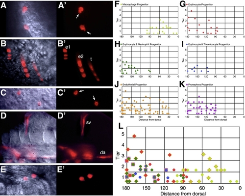
Hematopoietic Progenitors Originate from Both the Dorsal and Ventral Gastrula
(A–E) Examples of individual derivatives. (A) Macrophages, (B) erythrocytes and thrombocytes, (C) neutrophils, (D) endothelial cells, and (E) pronephric cells. da, dorsal aorta; e, erythrocytes; sv, segmental vein; t, thrombocytes. Arrows indicate cellular protrusions.
(F–L) 6 hr gastrula fate maps. Clones that included (F) macrophage cells, (G) just erythrocyte cells, (H) both erythrocyte and neutrophil cells, (I) both erythrocyte and thrombocyte cells, (J) endothelial cells, and (K) pronephric cells. (L) Summary hematopoietic fate map. Superimposed symbols show individual clones that gave rise to multiple fates. The half-green/half-blue symbol indicates the four clones that included all three ventral-derived blood fates (erythrocytes, neutrophils, and thrombocytes). Some of the blood clones depicted on the fate maps were also later verified by in situ hybridization.

Expression Data

Expression Detail
Antibody Labeling
Phenotype Data

Phenotype Detail
Acknowledgments
This image is the copyrighted work of the attributed author or publisher, and ZFIN has permission only to display this image to its users. Additional permissions should be obtained from the applicable author or publisher of the image.

Reprinted from Developmental Cell, 16(5), Warga, R.M., Kane, D.A., and Ho, R.K., Fate mapping embryonic blood in zebrafish: multi- and unipotential lineages are segregated at gastrulation, 744-755, Copyright (2009) with permission from Elsevier. Full text @ Dev. Cell