Fig. 11
- ID
- ZDB-FIG-090515-14
- Publication
- Easter et al., 1996 - The development of vision in the zebrafish (Danio rerio)
- Other Figures
- All Figure Page
- Back to All Figure Page
|
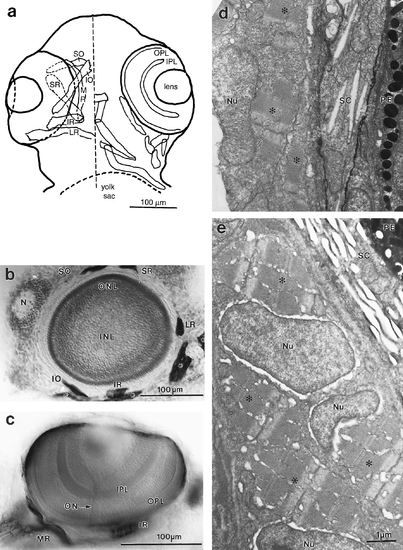
Extraocular muscles. (a) Camera lucida drawing of ventral view of the head of 96 hpf fish which had been reacted against the ZM-1 antibody. To the right of the midline (dashed line), the eye with its lens and two plexiform layers is indicated, as are the head muscles not associated with the eye. To the left, the six extraocular muscles are shown, with the convention that more ventral structures are outlined in solid, and masked outlines (of eye or muscles) are dashed. The six muscles are the superior oblique (SO), the inferior oblique (IO), the medial rectus (MR), the superior rectus (SR), the inferior rectus (IR), and the lateral rectus (LR). (b) Parasagittal (equatorial) section of a 96 hpf eye, dorsal up, rostral left, with approximately transverse sections of five of the six extraocular muscles. Only the medial rectus is missing, because it inserts more medially than the others. The nose (N) is indicated, as are the muscles not associated with the eye (*). (c) Optical section in the horizontal plane of a whole-mounted 96 hpf fish, lateral up, rostral left, showing the belly of the inferior rectus and the insertion of the medial rectus. The plexiform layers and the intraretinal optic nerve (ON) are also shown. (d, e) Electron micrographs of oblique sections through extraocular muscles in 72 and 96 hpf fish, respectively. The scale bar in e applies to both. The melanin in the pigmented epithelium (PE) and the reflective plates in the sclera (SC) provide landmarks for the edge of the eye. The nuclei (Nu) and myofibrils (*) are indicated. The older muscle is larger and more fully packed with myofibrils than the younger. |
Reprinted from Developmental Biology, 180, Easter, S.S., Jr. and Nicola, G.N., The development of vision in the zebrafish (Danio rerio), 646-663, Copyright (1996) with permission from Elsevier. Full text @ Dev. Biol.

