
Fig. 2
- ID
- ZDB-FIG-090511-12
- Publication
- Webb et al., 1997 - Localized calcium transients accompany furrow positioning, propagation, and deepening during the early cleavage period of zebrafish embryos
- Other Figures
- All Figure Page
- Back to All Figure Page
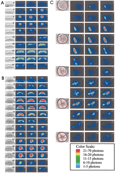
Representative (A) facial, (B) axial, and (C) top views of three f-aequorin-loaded zebrafish embryos demonstrating the changes in intracellular free calcium during the first and second cell division cycles. The photon images (colored panels) represent 30 s of accumulated luminescence with a 30-s gap between each image. Corresponding bright-field images were grabbed just after the preceding photon image. In the case of (A and B), this was every 3 min, while in the case of (C), every 9 min. In (C), for the sake of clarity, the outline of the blastodisc and locations of the furrows are outlined in red. Scale bar is 200 μm. |
Reprinted from Developmental Biology, 192, Webb, S.E., K.W. Lee, E. Karplus, and A.L. Miller, Localized calcium transients accompany furrow positioning, propagation, and deepening during the early cleavage period of zebrafish embryos, 78-92, Copyright (1997) with permission from Elsevier. Full text @ Dev. Biol.