
FIGURE
Fig. 3
- ID
- ZDB-FIG-090506-33
- Publication
- Waxman et al., 2009 - Increased Hox activity mimics the teratogenic effects of excess retinoic acid signaling
- Other Figures
- All Figure Page
- Back to All Figure Page
Fig. 3
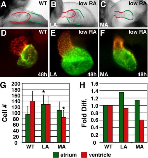
Excess RA signaling independently affects atrial and ventricular cell number. A-C: Lateral views of hearts at 48 hpf; red and green outlines indicate ventricle and atrium, respectively. D-F: Frontal views of Tg(cmlc2:DsRed2-nuc) (Mably et al.,[2003]) hearts, displaying nuclear DsRed in all cardiomyocytes, with Amhc immunofluorescence (green). G: Mean (±SD) number of atrial and ventricular cardiomyocytes. WT, n = 22; LA, n = 45; MA, n = 26. H: Fold difference of the means in G. Asterisks, statistically significant differences from WT (P < 0.005, Student's t-test). |
Expression Data
Expression Detail
Antibody Labeling
Phenotype Data
Phenotype Detail
Acknowledgments
This image is the copyrighted work of the attributed author or publisher, and
ZFIN has permission only to display this image to its users.
Additional permissions should be obtained from the applicable author or publisher of the image.
Full text @ Dev. Dyn.