
FIGURE
Fig. 5
- ID
- ZDB-FIG-090427-7
- Publication
- Wang et al., 2009 - Identification of wnt-responsive cells in the zebrafish hypothalamus
- Other Figures
- All Figure Page
- Back to All Figure Page
Fig. 5
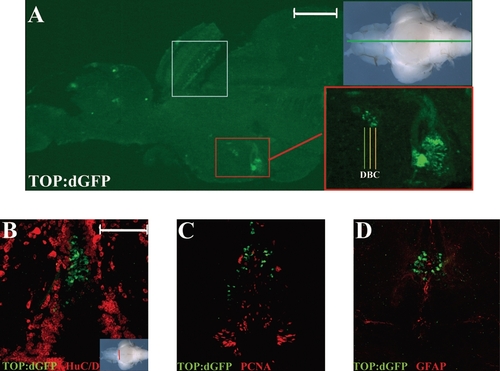
Immunohistochemical identification of Wnt-responsive and Lef1-expressing cells in the adult hypothalamus. (A) GFP antibody staining on a sagittal section through the midline of a TOP:dGFP adult brain. Specific expression of GFP is observed in the optic tectum (white box) and the periventricular hypothalamus (red box). (B–D) Twenty-five-micron hypothalamic cross sections immunostained for the markers listed in each panel. GFP-positive cells do not express HuC/D, PCNA, or GFAP. Scale bars: (A) 300 μm; (B) 100 μm. |
Expression Data
Expression Detail
Antibody Labeling
Phenotype Data
Phenotype Detail
Acknowledgments
This image is the copyrighted work of the attributed author or publisher, and
ZFIN has permission only to display this image to its users.
Additional permissions should be obtained from the applicable author or publisher of the image.
Full text @ Zebrafish